Laboratory of Volker H. Haase
Changes in tissue oxygen levels occur under pathological conditions and physiologically during development. The laboratory of Professor Volker H. Haase studies hypoxia response pathways and their therapeutic applications in erythropoiesis and iron metabolism, chronic kidney injury and ischemic pre-conditioning, inflammation, kidney development and tumorigenesis.
A major focus of the lab is on the interplay between hypoxic signaling, metabolism and cellular differentiation and its regulation by the prolyl hydroxylase domain (PHD) / hypoxia-inducible factor (HIF) / von Hippel-Lindau tumor suppressor (VHL) signaling axis. Haase group members take advantage of powerful cutting-edge mouse genetics, biochemical, metabolomic and single cell approaches to study oxygen and mitochondrial metabolism in kidney, urologic and other diseases. Click on links for information about career opportunities in the Haase lab and recent publications.
Introduction to HIF and PHD oxygen sensors
The PHD / HIF axis is a critically important oxygen-sensing pathway that mediates tissue adaptation to low oxygen environments primarily via the transcriptional regulation of gene expression. The laboratory of Volker H. Haase investigates the key components of this pathway and their regulation by hypoxia. The identification of oxygen- and iron-dependent PHD enzymes as key regulators of HIF has led to the development of novel therapeutic agents that are currently in clinical development for the treatment of anemia associated with chronic kidney disease.
Hypoxia-inducible factor
HIFs are basic helix-loop-helix transcription factors and members of the PAS (PER/aryl hydrocarbon receptor nuclear translocator (ARNT)/single minded (SIM)) family of transcription factors. They consist of an oxygen-sensitive α-subunit and a constitutively expressed β-subunit, which is often referred to as the aryl hydrocarbon receptor nuclear translocator (ARNT).
Three HIF-α-subunits have been identified, HIF-1α, HIF-2α (also known as EPAS1) and HIF-3α. HIF-1 and HIF-2 are the most extensively studied HIF transcription factors and facilitate oxygen delivery and adaptation to hypoxia by regulating a wide spectrum of cellular and tissue hypoxia responses. These include the stimulation of red blood cell (rbc) production and angiogenesis, the induction of glycolysis, reductions in fat and mitochondrial metabolism, as well as alterations in cardiovascular function.
HIF-α-subunits, although continuously synthesized, are rapidly degraded in the presence of molecular oxygen. When cells experience hypoxia HIF-α is no longer degraded and translocates to the nucleus where it forms a heterodimer with HIF-β and activates gene transcription.
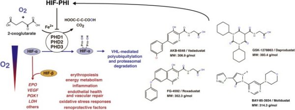
(click on image to enlarge)
Schematic overview of the PHD/HIF pathway. Although the oxygen-sensitive α-subunit of HIF is constitutively synthesized, it is rapidly degraded under normoxic conditions. Under hypoxia, however, cellular HIF-α levels build up and HIF-α translocates to the nucleus, where it forms a heterodimer with HIF-β. Proteasomal degradation of HIF-α is mediated by the pVHL-E3-ubiquitin ligase complex and requires HIF-α prolyl-4-hydroxylation by oxygen- and iron-dependent PHD dioxygenases (PHD1-3). The decarboxylation of 2-oxoglurate (2OG) produces hydroxylated HIF-α, succinate and CO2. PHD or VHL inhibition results in increased transcription of HIF-regulated genes such as vascular endothelial growth factor (VEGF), erythropoietin (EPO), phosphoglycerate kinase 1 (PGK1), lactate dehydrogenase (LDH) and other genes involved in the regulation of hypoxia responses, including cellular metabolism and mitochondrial function, inflammation, vascular function and oxidative stress and other responses. Shown are also the chemical structures of PHD inhibitors (PHI) that are in phase III clinical development and are capable of effectively stimulating the production of endogenous EPO in patients with chronic kidney disease.
PHD oxygen sensors
Hydroxylation of specific proline residues is required for normoxic HIF-α degradation and is carried out by PHD1, PHD2 and PHD3, which function as the oxygen sensors of the HIF pathway. HIF-PHDs belong to a large family of 2-oxoglutarate (OG)-dependent dioxygenases. These enzymes utilize molecular oxygen for hydroxylation and thus couple oxygen, intermediary and amino acid metabolism to multiple cellular processes, which include HIF responses, collagen synthesis, fatty acid metabolism and the regulation of the epigenome. Any reduction in HIF-proline hydroxylation impairs HIF-α degradation and leads to the activation of HIF-mediated cellular hypoxia responses.
Structural analogs of 2OG that reversibly inhibit HIF-proline hydroxylation {HIF-prolyl hydroxylase inhibitors (HIF-PHIs)} have been shown to effectively stimulate HIF responses in the presence of normal oxygen levels and are now in clinical development for renal anemia therapy and other indications (click here for a recent review on this topic).